Váš nákupní košík je prázdný


Počítačová tomografie
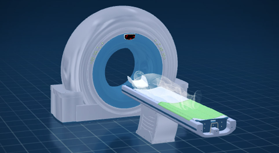
Pomocí této animace poznáme strukturu a fungování počítačové tomografie.
Technologie
9. – 12. ročník
Klíčová slova
mozaLink
/Weblink
Scénky
Počítačová tomografie (CT)
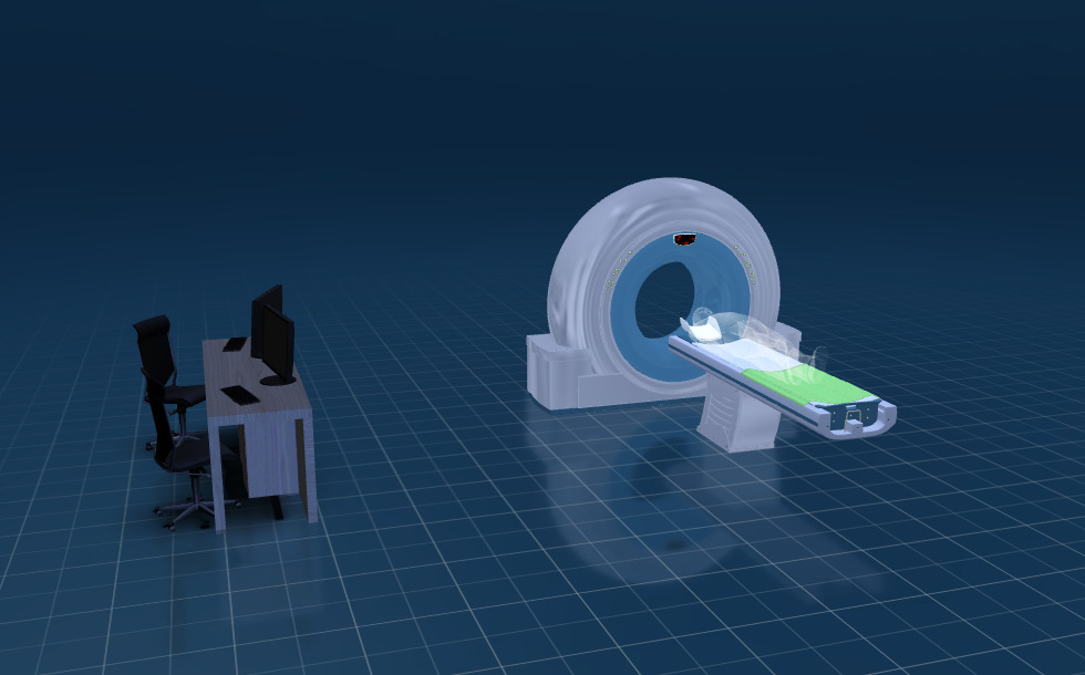
- počítač na zpracování údajů
- gantry
- automatická postel
- generátor
Conventional X-ray images show the extent to which a beam of X-rays is absorbed while passing through tissues in the body. Tissues that absorb a great amount of radiation, such as bones, are well-defined in X-ray images. Air-filled spaces are also easily identified because they do not absorb X-rays. X-ray images represent overlapping tissues in their totality, making it challenging to examine internal organs, soft tissues and the brain. However, the diagnosis of numerous injuries and diseases requires sharp images of the tissue layers not visible in a conventional radiographic image.
Computed tomography is an improved version of conventional radiography, developed to map these areas. It was developed by scientists Allan M. Cormack and Godfrey N. Hounsfield. This achievement earned them the Nobel Prize in Physiology or Medicine in 1979.
Konstrukce
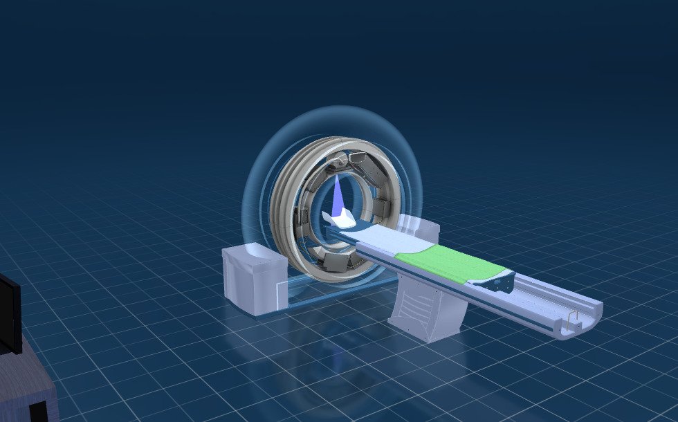
- skener
- detektor
- rentgenka
- rentgenové záření
The CT scan is carried out in the ring-shaped frame of the device, called the gantry, which houses the X-ray tube that emits the beams, as well as the detector that captures radiation. X-ray tubes found in CT devices produce a narrow, fan-shaped beam of X-rays that penetrates through the body in a single plane.
Fungování
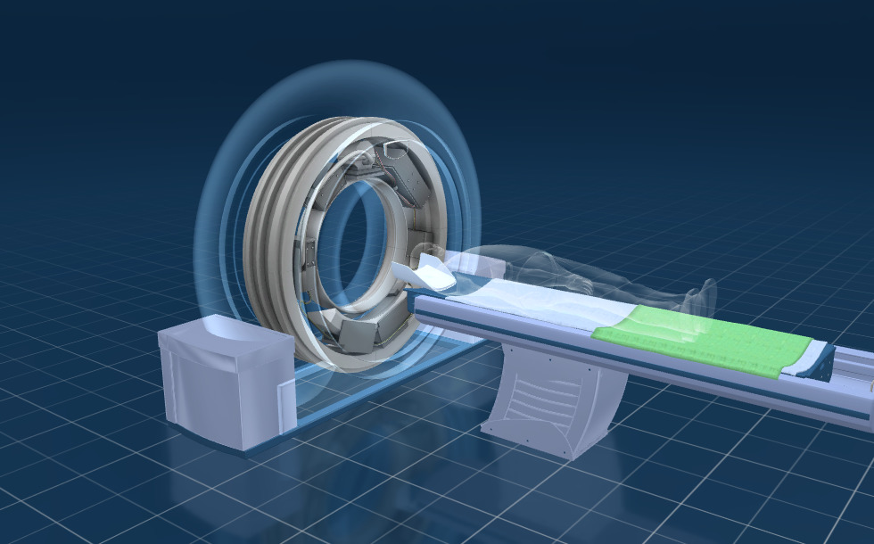
The word tomography derives from the Greek words 'tomos' (slice) and 'grapho' (to write). Computed tomography is a technique used to obtain digital cross-sectional images ('slices') of the body.
The CT scanner measures the amount of radiation absorbed in each area. The body is scanned from several directions using a rotating ring that contains an X-ray tube and detectors on the opposite side. The multiple measurements performed during one rotation form an intensity graph that helps map the given plane in detail. When a 'slice' is completed, the motorised bed moves slightly and another cross-sectional scan is taken. The intensity graphs of the slices are then assembled by the device, forming a 3D image of the body.
How much radiation is a patient exposed to during a CT examination? The average natural background radiation on the Earth is about 3 millisieverts per year, originating from cosmic rays and the decay of radioactive materials found in the soil and rocks. A head CT exposes the body to a radiation dose of 2 millisieverts, while chest, abdominal and spinal CT scans the body receives 7, 8 and 6 millisieverts of radiation, respectively. Due to the relatively high radiation exposure, this diagnostic method is recommended only in medically justified cases.
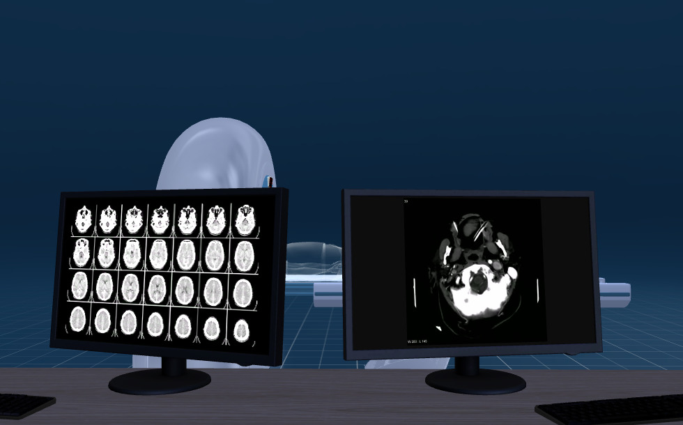
Zobrazování
Certain tissues cannot be distinguished in conventional X-ray images, as they absorb nearly the same amount of X-rays. In addition, they can also be covered by denser tissues, such as bones, which block the passage of the X-ray beam.
As the skull absorbs X-rays to a great extent, the brain cannot be examined with conventional X-ray machines. However, using computed tomography, high-detail images can be taken. This new method allowed radiographic imaging of the brain for the first time.
In CT scans, the areas that absorb radiation to the greatest extent appear white, while those that do not absorb x-rays show as black. Bones are seen as white, air appears black, soft tissues are indicated by shades of light grey and fat is shown in dark grey.
Computed tomography is commonly used to examine the head, the chest, the abdominal organs and the blood vessels. With the help of this imaging technique, it is possible to diagnose medical conditions such as strokes, brain tumours and arteriosclerosis, as well as abdominal tumours, lung disorders or coronary artery diseases. A contrast agent is often administered to improve the visibility of the organs and tissues.
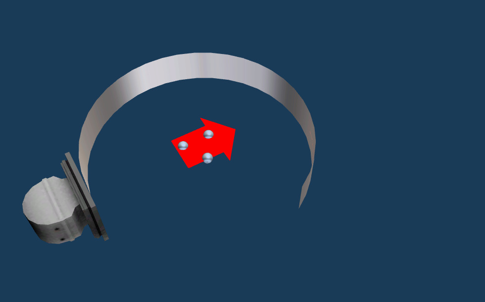
Princip zobrazování
- počítačové zpracování dat
- detektor
- zkoumaný objekt
- projekce zkoumaného objektu
Each image is taken from a slightly different angle so the absorption of X-rays can be seen from every direction. The absorption data obtained by the rotation of the X-ray source is processed and transformed into a cross-sectional image, that is, a 'slice' of the body.
Vyprávění
Computed tomography (CT) is a diagnostic imaging method that uses X-rays to map the internal spatial structure of the human body without making any surgical incisions. CT is an improved version of conventional radiography developed by scientists Allan M. Cormack and Godfrey N. Hounsfield. This achievement earned them the Nobel Prize in Physiology or Medicine in 1979.
The CT scan is carried out in the ring-shaped frame of the device, called the gantry, which houses the X-ray tube that emits the beams as well, as the detector that captures radiation. X-ray tubes found in CT devices produce a narrow, fan-shaped beam of X-rays that penetrates through the body in a single plane.
The CT scanner measures the amount of radiation absorbed in each area. The body is scanned from several directions using a rotating ring that contains an X-ray tube and detectors on the opposite side. The multiple measurements performed during one rotation form an intensity graph that helps map the given plane in detail. When a 'slice' is completed, the motorised bed moves slightly and another cross-sectional scan is taken. The intensity graphs of the slices are then assembled by the device, forming a 3D image of the body.
Computed tomography is commonly used to examine the head, the chest, the abdominal organs and the blood vessels. With the help of this imaging technique, it is possible to diagnose medical conditions such as strokes, brain tumours and arteriosclerosis, as well as abdominal tumours, lung disorders or coronary artery diseases. A contrast agent is often administered to improve the visibility of the organs and tissues. The examination takes only 5–20 minutes, however, as the radiation produced during the CT scan is much higher than the average natural background radiation, this diagnostic method is recommended only in medically justified cases.
Each image is taken from a slightly different angle so the absorption of X-rays can be seen from every direction. The absorption data obtained by the rotation of the X-ray source is processed and transformed into a cross-sectional image, that is, a 'slice' of the body.